- +91 83103 67685
- info@drameetoswal.com
- Basavanagudi
A revolutionary imaging tool that gives a clear understanding of your blood flow condition.

Overview
Intravascular Imaging is a technique that helps your cardiologist to get inside the artery. This is done with a camera-like device that provides a real-time view of the artery of heart. It gives information like severity of blockage, abnormalities like calcium, dissection and thrombus. It can also be used in planning for the insertion of stents.
During the procedure, the cardiologist will make a small incision in your groin to gain access to a large artery. Your cardiologist, with a slim guide wire, will gently advance the wire to the arteries of your heart. There are three ways that this technique is executed:

If you have significant blockages or leakages or narrowing that can hinder the blood flow, that could mean you may need an angioplasty. It is indicated in patients who needs complex angioplast like CTO intervention, Calcified coronary arteries, Bifurcation lesions & multi vessel disease. If a coronary artery becomes partially blocked, it can hinder the oxygen supply to areas of the heart. This can result in a heart attack. Some of the common reasons to see a doctor can be:
If you have any such symptoms, the doctor might suggest an angioplasty. This would subsequently mean, that you have to undertake Intravascular Imaging in Basavanagudi.
The doctor might suggest angioplasty if you have the following symptoms:
This would subsequently mean that you have to undertake Intravascular Imaging.
It is a minimally invasive procedure used to get images from inside the arteries. This is usually done under conscious or minor sedation. During the process, your cardiologist will make a small incision in your groin to get a large artery. They will insert a very thin guide wire topped with a pressure sensor. Your cardiologist will gently advance the wire to the arteries of your heart. You may feel mild pressure or a slight burning sensation in your chest during this procedure
It enables a physician to get inside the artery with a camera-like device. It can quantify the percentage of narrowing and give insight into the nature of the plaque. This helps to calculate the extent of the medical care to be administered.
Helps to produce calcifications as bright images with shadowing behind them. Images for the blood vessel wall inner lining, atheromatous disease within the wall, connective tissues, and blood and healthy muscular tissue.
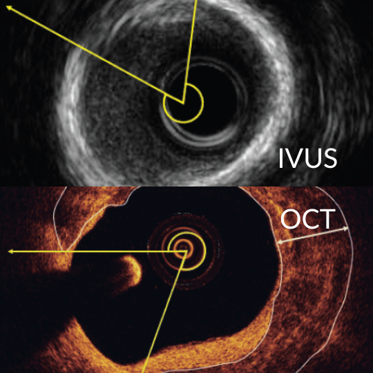
IVUS uses ultrasound and OCT uses infrared lights, but both methods use the same probing technique each has its own advantage for specific type of procedure planned.
FFR uses a pressure sensor probe to calculate blood pressure difference in the artery while IVUS is used to get cross-sectional images with ultrasound. They both are used as compliments for the imaging process.
Subscribe our newsletter for latest information in the field of cardiology
WhatsApp us