- +91 83103 67685
- info@drameetoswal.com
- Basavanagudi
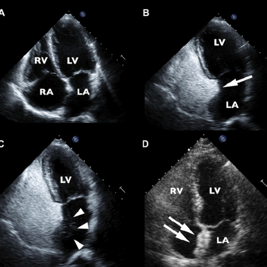
Contrast Echocardiography in Bangalore | A newly developed type of echocardiography that gives more clarity to medical images produced

A harmless contrast agent is injected into your bloodstream before carrying out an echocardiogram. This substance helps to create clearer images on the screen because of its high reactivity to sound. Blood usually appears just “black” in medical imaging, hence it becomes difficult to visualize aptly what problems there might be. Contrast is used in an echocardiogram to see the flow of blood and the heart muscle clearly.

Contrast Echocardiography in Bangalore | Contrast Echocardiography can help detect
certain heart conditions by checking the structure of the heart and surrounding
blood vessels, analysing blood flow, and assessing the pumping chambers of the
heart. It can be used to find out about the following:
Contrast echocardiography can also help your cardiologist decide on the best treatment for these conditions.
A contrast echo is consequently done when normal echocardiogram images are not of high enough quality to see all the details of the heart and its major blood vessels. From beginning to end, the test takes about 30 minutes. An echocardiogram lets your cardiologist see how well the heart muscle is working, see how the valves are functioning, and check for abnormal connections in the heart chambers.
The process is similar to that of normal echocardiography, with only some minor differences. The procedure of contrast echocardiography has the following steps:
Saline (sterile salt water) and Definity are the two common contrast solutions which may be used. Each can showcase blood flow through the heart and improve the image of the heart that is produced.
Saline is one of the contrasting agents that are used. It is firstly shaken to create bubbles which are harmless, to be injected into the IV.
Definity is another used this is also injected into the IV during the echocardiogram. And its presence can be clearly seen in the heart.
No, it is a completely safe process. The contrasting agents are tried and tested to prove that they are harmless to the heart and body.
It usually takes up to 30-60 minutes of time.
Subscribe our newsletter for latest information in the field of cardiology
WhatsApp us